

Can Damaged Dna Be Repaired
Abstract
Aging is a complex process that results in loss of the ability to reattain homeostasis following stress, leading, thereby, to increased adventure of morbidity and bloodshed. Many factors contribute to crumbling, such as the time-dependent aggregating of macromolecular damage, including DNA damage. The integrity of the nuclear genome is essential for cellular, tissue, and organismal health. DNA damage is a abiding threat because nucleic acids are chemically unstable under physiological conditions and vulnerable to attack by endogenous and environmental factors. To combat this, all organisms possess highly conserved mechanisms to observe and repair Deoxyribonucleic acid impairment. Persistent Deoxyribonucleic acid impairment (genotoxic stress) triggers signaling cascades that drive cells into apoptosis or senescence to avoid replicating a damaged genome. The drawback is that these cancer avoidance mechanisms promote aging. Here, nosotros review evidence that DNA damage plays a causal role in aging. We as well provide evidence that genotoxic stress is linked to other cellular processes implicated as drivers of aging, including mitochondrial and metabolic dysfunction, altered proteostasis and inflammation. These links between damage to the genetic code and other pillars of crumbling support the notion that DNA impairment could be the root of aging.
Introduction
Aging is a multifactorial procedure that results in increased risk of a myriad of chronic diseases. Existence elderly is the greatest take a chance factor, by orders of magnitude, for cancer, osteoporosis, cardiovascular affliction, dementia and most other degenerative diseases (Kirkwood, 2005). While no single mechanism or pathway fully accounts for age-associated functional reject, i prevailing theory is that macromolecular damage, accumulating over fourth dimension, plays a causal office in driving aging. Nigh macromolecules in the cell when damaged are simply degraded and replaced. In contrast, the nuclear genome, which is the blueprint for all cellular functions, has dedicated and energetically costly repair mechanisms to rapidly correct Dna damage. This intimates that Deoxyribonucleic acid harm is a specially hazardous blazon of macromolecular harm and therefore probable to be deleterious to cellular homeostasis.
Maintaining genome stability is a continuous process. Deoxyribonucleic acids are chemically unstable nether physiological conditions (aqueous, oxygen-rich, and pH 7.iv) (Lindahl, 1993). Dna is too vulnerable to chemical attack by electrophiles and complimentary radicals. While exogenous sources of genotoxic stress can be quite potent, endogenous threats are constant and relentless (Tabular array 1). The about mutual DNA lesion is hydrolytic cleavage of the glycosidic bail between the DNA base and sugar phosphate group, leading to abasic sites. Hydrolytic deamination of the DNA bases is too common. Products of normal cellular metabolism can cause oxidation, nitrosylation, and alkylation of the Deoxyribonucleic acid bases (De Bont and van Larebeke, 2004). Breaks in the phosphate deoxyribose backbone arise as a consequence of loftier free energy radiation or during Deoxyribonucleic acid metabolism (replication, decatenation). Spontaneous DNA harm occurs on the order of 104–x5 events per cell per 24-hour interval (Lindahl, 1993; De Bont and van Larebeke, 2004).
Estimated frequencies of Deoxyribonucleic acid lesions caused by endogenous and common environmental sources of DNA damage.
Adapted from Friedberg, 2006; Lindahl, 1993; Sander et al., 2005; Sears and Turchi, 2022; Mouret et al., 2006.
| Endogenous DNA adducts | |||||||||
|---|---|---|---|---|---|---|---|---|---|
| Dna lesion | DSB | Cytosine deamination | Cyclopurine adducts | Depyrimidination | 8-oxoG | Malondialdehyde adducts | Alkylation adducts | Depurination | SSB |
| Frequency per cell per day | 101 | 102 | x2 | 102 | 10three | 10iii | 10three | 104 | 104 |
| DNA adducts caused by environmental exposures | |||||||||
| Genotoxin | Sunlight | Background radiation | Ionizing radiation therapy | Oxaliplatin cancer therapy | |||||
| Lesion | Photodimers | Damaged bases | SSB | DSB | Damaged bases | SSB | Intra- and interstrand crosslinks | ||
| Frequency per cell per 24-hour interval | ten2 in skin cells only | 10 | two–v | 0.25 | ten3 | 103 | 10three | ||
DNA is also susceptible to impairment by environmental factors such as ultraviolet (UV), ionizing radiations, and alkylating agents used to care for proliferative disorders similar cancer (Table one). Notably, even when exogenous genotoxin exposure is instigated with the purpose of driving cell death (east.chiliad., in cancer therapy) adduct burdens are well beneath the incidence of endogenous damage (Jackson and Loeb, 2001). Fortunately, all organisms take robust mechanisms to sense all types of DNA damage, delay genome replication (if needed), signal for repair, and right or tolerate the large number of genomic insults that occur on a daily footing (Hoeijmakers, 2009). DNA harm that is not repaired in a timely manner or is too egregious to be repaired induces signaling events that atomic number 82 to 1 of many cell fates, 1 of which, senescence, plays a causal role in aging.
Conceptually, could DNA damage bulldoze aging?
Why? and how? organisms historic period remain challenging questions. Why 1 ages interrogates the reasons. How ane ages interrogates the method. The combative pleiotropic theory of aging provides a genetic solution to why we age, posing that genes that provide an advantage during reproductive life are disadvantageous post reproduction, nonetheless these genes cannot exist selected against (Kirkwood, 2005). Equally an example of antagonistic pleiotropy, activation of the Deoxyribonucleic acid impairment response (DDR) is critical for preventing cancer; however, chronic activation of the DDR is thought to drive the accumulation of senescent cells and chronic sterile inflammation in old age, equally described in more detail beneath. The pillars of aging (Kennedy et al., 2022) describe the method by which (or how) we age: loss of or impaired, mitochondrial integrity and function, metabolism, stem prison cell office, proteostasis, food sensing, accommodation to stress, autophagic flux, epigenetic control, and an aggregating of damaged cellular macromolecules. This includes damage to the nuclear and mitochondrial genomes. Yet, it has proven challenging to constitute the pillars of aging as truthful causes of crumbling rather than just consequences of crumbling.
If DNA impairment drives aging, mechanistically how does it do and then? Through activating signaling responses (d'Adda di Fagagna et al., 2003), blocking transcription (Vermeij et al., 2022) and other DNA metabolism, altering the epigenome (Oberdoerffer et al., 2008), mutagenesis (Vijg, 2022), triggering cells senescence or apoptosis? Deoxyribonucleic acid harm occurs stochastically but the amount and types of Deoxyribonucleic acid damage one experiences is influenced by the expression of genes encoding antioxidant enzymes, genes linked to energetics and mitochondrial function, and a myriad of other factors such every bit histones, methylases, sirtuins, transcription, and replication factors. Every aspect of how DNA damage might drive aging is also genetically determined via the cellular response to Deoxyribonucleic acid impairment. The somewhat surprising finding is that DNA damage has far-reaching effects on many aspects of cellular metabolism tied to aging, the then-chosen pillars of aging (Kennedy et al., 2022). This suggests that aging might be driven by many types of cellular impairment notwithstanding does not occur until one reaches a state where multiple aspects of cell biology are perturbed, for case, genome integrity, proteostasis, and mitochondrial function.
DDR and cell fate decisions
Once DNA damage is recognized in the nuclear genome, bulky adducts, small miscoding lesions, single-strand breaks, or non-circuitous double-strand breaks (DSBs) can exist direct repaired by nucleotide excision repair (NER), base excision repair (BER), and non-homologous end-joining (NHEJ), respectively. If replication forks or transcription complexes see polymerase-blocking lesions (Vermeij et al., 2022), this can pb to the germination of a DSB or R-loop (Tresini et al., 2022), which potently activate signaling events that halt jail cell bike progression and promote repair. If the damage signals persist, so the jail cell selects a fate that avoids replicating a damaged genome and mutagenesis at all cost, by activating events that lead to cell death (apoptosis) or irreversible jail cell wheel arrest (senescence) (Figure ane). DNA harm signaling begins with the MRE11-RAD50-NBS1 (MRN) complex activating the phosphatidylinositol 3-kinase-like kinases (PIKKs) clutter-telangiectasia mutated (ATM), ATM-related kinase (ATR) and/or related PIKK (Thompson, 2022). ATM is activated primarily by DNA DSBs while ATR is primarily involved in the response to stalled replication forks, although overlap occurs. Initially ATM and ATR piece of work with checkpoint mediator proteins similar MDC1, 53BP1 and BRCA1, TOPBP1, which are sensor proteins that bind to lesions and recruit other DDR factors. Harm recognition is followed by phosphorylation (and activation) of transducer kinases like checkpoint kinase 2 (CHK2) or checkpoint kinase 1 (CHK1), which amplify the ATM-ATR betoken. CHK2 activation leads to activation of p53, a primary regulator of the cellular response to genotoxic stress (Senturk and Manfredi, 2022). Sustained activation of these transducer proteins leads to phosphorylation of several Dna repair and cell bicycle checkpoint proteins (Thompson, 2022; Cheng and Chen, 2010). p53 can also be activated independently of ATM. p14/ARF inhibits MDM2–p53 interaction, resulting in the stabilization of p53 (Senturk and Manfredi, 2022; Meek and Anderson, 2009). p53 tin mount a bimodal response to stress where it functions to actuate apoptosis via diverse transcriptional targets like Bax, Puma, and Noxa, or promote transient or prolonged jail cell wheel abort through transcriptional activation of the cyclin-dependent kinase inhibitor p21CIP1 (Reinhardt and Schumacher, 2022). A myriad of factors determines the extent of p53 activation in response to genotoxic stress, and more than factors likely will be discovered.
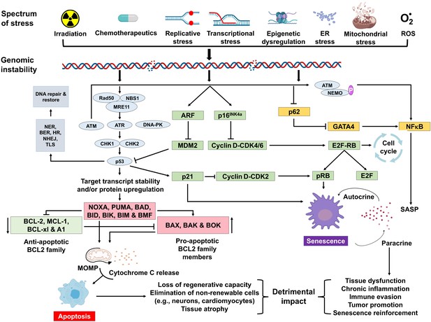
Schematic representation of signaling events within a cell that enable DNA harm to promote aging.
Depicted are diverse stressors that tin can lead to genome instability and activation of the DNA damage response (DDR). The DDR (lite blue) leads to jail cell bike arrest (green). If signaling persists, apoptosis or senescence ensues. Senescence tin can affect neighboring, undamaged cells.
A variety of types of stress can activate the INK4a/ARF locus in cells. The gene products of the locus—p16INK4a and ARF—arrest cell progression past interim on the retinoblastoma protein and p53 (Senturk and Manfredi, 2022). p16INK4a inhibits CDK4 from binding to cyclin D, thereby preventing phosphorylation of RB. Hypo-phosphorylated retinoblastoma (RB) protein represses E2F-dependent gene expression, blocking G1/Due south prison cell cycle progression. Free E2F activates many genes involved in mitosis. While sensing Deoxyribonucleic acid damage and responding to it is a positive attribute, persistent Deoxyribonucleic acid damage signaling can have quite deleterious furnishings. It can lead to chronic activation of p53 and other response pathways (stress response, pro- and anti-apoptotic, pro-inflammatory, etc.), which affect many aspects of cellular function and impact jail cell fate. Definitive cognition well-nigh which factors determine the selection of jail cell fate choice remains elusive. However, information technology seems likely that these factors may vary with the cell blazon, the level, and the elapsing of genotoxic stress.
Cellular senescence and the secretory phenotype
Leonard Hayflick and Paul Moorhead divers cellular senescence as the proliferative abort that occurs in normal cells after a number of cell divisions (Hayflick and Moorhead, 1961; Hayflick, 1965). The commencement molecular explanation for senescence was the progressive telomere shorting that occurs with every cell partition termed telomere-initiated cellular senescence (Harley et al., 1990). Many cells undergo senescence independently of telomere shorting due to replicative, mitochondrial, oxidative, metabolic, or genotoxic stress (Parrinello et al., 2003; Christoffersen et al., 2010; Correia-Melo et al., 2022; Nair et al., 2022). Senescent cells actively repress cell cycle progression preventing the replication of a damaged genome. Senescent cells have altered metabolism, morphology, and secretory profile called the senescence-associated secretory phenotype (SASP). The SASP includes pro-inflammatory cytokines, chemokines, and proteases (Coppé et al., 2008) and is heterogenous depending on the jail cell type and the inducer of senescence (Basisty et al., 2022). The SASP is idea to instigate immune clearance of the damaged/stressed cells (Tasdemir et al., 2022), but the immediate negative upshot is the potential to drive local tissue damage and systemic chronic sterile inflammation (Franceschi and Campisi, 2022).
Ane of the challenges of studying senescence is the absence of a specific marking to place senescent cells. Therefore, multiple endpoints must exist measured simultaneously to identify them (Gorgoulis et al., 2022; Sharpless and Sherr, 2022). The most widely used marker is measurement of senescence-associated β-galactosidase (SA-β-gal) activity (Itahana et al., 2007). Other markers associated with cellular senescence include absence of proliferation markers (east.g., Ki-67), increased expression of the cyclin-dependent kinase inhibitors p16INK4a and p21CIP1, persistent activation of the DDR (such as p53, ATM, ATR, ɣH2AX, telomere dysfunction-induced foci [TIF], senescence-associated heterochromatin foci [SAHF], and DNA segments with chromatin alterations reinforcing senescence [DNA-SCARS]), reduced lamin B1 expression, increased HMGB1 and SASP factors (such as IL-1β, IL-6, IL-x, MCP-i, and TNFα) (Coppé et al., 2010; Lawless et al., 2010; Serrano et al., 1997; Takai et al., 2003; Matjusaitis et al., 2022; Rodier et al., 2022).
Cellular senescence plays a disquisitional role in preventing tumorigenesis, tissue repair, and wound healing, promoting insulin secretion and mammalian development (Kuilman et al., 2008; Krizhanovsky et al., 2008; Demaria et al., 2022; Storer et al., 2022; Helman et al., 2022). Notwithstanding, time-dependent accumulation of senescent cells, likely through decreased immunoclearance, occurs in virtually all vertebrates (Jeyapalan et al., 2007; Liu et al., 2009) and has been definitively shown to drive aging (Baker et al., 2022), rather than simply occur with aging. Senescent cells besides contribute to age-related diseases such as atherosclerosis (Roos et al., 2022), osteoporosis (Chandra et al., 2022), nonalcoholic fatty liver disease (Ogrodnik et al., 2022), cancer (Alimirah et al., 2022), neurodegenerative diseases (Chinta et al., 2022; Zhang et al., 2022), and numerous other historic period-related conditions (Kirkland and Tchkonia, 2022). Interestingly, NF-κB is the transcription cistron that is nigh activated with aging (Adler et al., 2007). NF-κB signaling is the major signaling pathway that stimulates SASP (Tilstra et al., 2022). Dna damage activates NF-κB signaling via an ATM-NEMO-dependent regulation of an upstream kinase (Miyamoto, 2022; Effigy i). Dna damage can also lead to NF-κB activation via stabilization of GATA4 (Kang et al., 2022). Thus, genotoxic stress is a potent inducer of senescence and SASP, key drivers of aging and age-related disease.
Accelerated aging in genome instability syndromes
The mammalian genome encodes over 150 proteins directly responsible for safeguarding its integrity (Friedberg, 2006; Wood et al., 2005). These cistron products constantly monitor the quality and repair the nuclear genome (Sancar et al., 2004). Distinct DNA repair pathways cope with different types of DNA lesions: BER for small covalent additions to DNA bases, NER for bulky adducts that disrupt the DNA helix, interstrand crosslink (ICL) repair to remove covalent links between the two strands of Deoxyribonucleic acid, NHEJ to ligate broken DNA ends, mismatch repair to correct replication errors, homologous recombination to manage replication stress and DSBs not readily ligated. Inherited defects in each of the Deoxyribonucleic acid repair pathways are linked to distinct genome instability syndromes (Tabular array 2). Broadly, the syndromes are characterized by developmental defects, increased incidence of cancer and features of accelerated aging (Cleaver, 1968; Menck and Munford, 2022; Wood, 2022). Progeroid syndromes are diseases of dramatically accelerated crumbling and include Hutchinson-Gilford, Werner, and Cockayne syndromes (CS), as well as XFE progeroid syndrome, all of which are linked to genome instability (de Magalhães, 2005; Martin and Oshima, 2000; Burla et al., 2022). The syndromes provide an elegant yet tragic glimpse into the impact that DNA damage tin have on human health. Nigh syndromes were described well before DNA had been discovered or mechanisms of repair described.
Homo genome instability diseases with age-associated symptoms.
| Disease | Affected genome stability pathway | Mutated genes | Aging-associated symptoms | Ref(s) |
|---|---|---|---|---|
| Hutchinson-Guilford progeria syndrome | Chromatin organisation | LMNA | Baldness, atherosclerosis, arthritis, cardiovascular disease, lipodystrophy, osteoporosis, peel aging and cloudburst | Kudlow et al., 2007; Liu et al., 2005 |
| Nestor-Guillermo progeria syndrome | Chromatin organization | BANF1 | Baldness, atherosclerosis, arthritis, cardiovascular illness, lipodystrophy, osteoporosis, and pulmonary hypertension | Cabanillas et al., 2022; Loi et al., 2022 |
| Werner syndrome | Telomeric maintenance and replication stress | WRN | Alopecia, atherosclerosis, arthritis, cardiovascular illness, cataracts, diabetes, sarcopenia, and increased risk of cancer | Kudlow et al., 2007; Sugimoto, 2022 |
| Rothmund-Thomson syndrome | Deoxyribonucleic acid replication initiation | RECQL4 | Alopecia, cataracts, osteoporosis, peel atrophy, and increased chance of cancer | Croteau et al., 2022; Ghosh et al., 2022 |
| Bloom syndrome | DNA replication and recombination | BLM | Diabetes, pulmonary illness, increased risk of cancer | Hanada and Hickson, 2007; de Renty and Ellis, 2022 |
| XFE progeroid syndrome | NER, ICL, and DSB repair | ERCC4 | Anemia, cardiovascular disease, kidney disease, neurodegeneration, osteoporosis, sarcopenia, sensory loss, and pare cloudburst | Niedernhofer et al., 2006 |
| Xeroderma pigmentosum | NER and translesion DNA synthesis | XPA-K, XPV | Premature skin photoaging, neurodegeneration, and increased incidence of skin cancer | Lehmann et al., 2022; Kraemer and DiGiovanna, 2022 |
| Cockayne syndrome | Transcription-coupled NER | CSA, CSB, XPB, XPD, XPG | Ataxia, cataracts, muscle atrophy, and neurodegeneration | Nance and Drupe, 1992; Wilson et al., 2022 |
| Trichothiodystrophy | Transcription-coupled NER | TTDA, TTDN1, XPB, XPD | Premature bone marrow exhaustion and increased risk of cancer | Faghri et al., 2008; de Boer et al., 2002 |
| Fanconi anemia | ICL repair | FANCA-FANCW | Premature bone marrow exhaustion and increased risk of cancer | Ceccaldi et al., 2022; Nalepa and Clapp, 2022 |
| Ataxia telangiectasia | DNA impairment response | ATM | Premature bone marrow exhaustion, diabetes, and neurodegeneration | Rothblum-Oviatt et al., 2022 |
| Mandibular hypoplasia, deafness, progeroid features, lipodystrophy syndrome | Post-replication repair and translesion Deoxyribonucleic acid synthesis | POLD1 | Diabetes, lipodystrophy, osteoporosis, steatosis, sensory loss | Weedon et al., 2022 |
| Ruijs-Aalfs syndrome | Poly peptide-DNA crosslink repair | SPRTN | Alopecia, atherosclerosis, cataracts, diabetes, premature graying of hair, osteoporosis, sarcopenia, and increased risk of cancer | Lessel et al., 2022 |
| Alpers-Huttenlocher syndrome | Mitochondrial DNA replication and repair | POLG1 | Progressive neurodegeneration and liver illness | Nguyen et al., 2006 |
Children with Hutchinson-Gilford progeroid syndrome (HGPS) develop many features of premature aging in the first decade of life including alopecia, atherosclerosis, osteolysis, and lipodystrophy among others (Hennekam, 2006). The genetic cause for HGPS are mutations in the LMNA gene, which encodes critical components of the nuclear lamina (Eriksson et al., 2003; De Sandre-Giovannoli et al., 2003) causing pernicious alterations in nuclear architecture resulting in genome instability (Kudlow et al., 2007). Nestor-Guillermo progeroid syndrome, caused by mutations in the nuclear lamina gene BANF1, has characteristics of accelerated aging due to impaired chromatin organization (Cabanillas et al., 2022; Loi et al., 2022).
Mutations in DNA helicases are linked to multiple diseases of accelerated aging. Mutations in three of the RECQ family of helicases (BLM, RECQL4, and WRN) cause progeroid syndromes (Uchiumi et al., 2022). Mutations in WRN, a factor that encodes a helicase responsible for managing replication stress and telomere stability, cause Werner syndrome (WS) (Kudlow et al., 2007). WS patients feel growth retardation, premature hair graying, lipodystrophy, likewise as premature onset of multiple age-related diseases including arteriosclerosis, cancer, blazon 2 diabetes mellitus, and osteoporosis (Sugimoto, 2022). Rothmund-Thomson syndrome (RTS) is caused by mutations in RECQL4, encoding a helicase that participates in DNA replication and repair. RTS patients experience juvenile cataracts, epidermal cloudburst, and increased cancer incidence (Croteau et al., 2022; Ghosh et al., 2022). Bloom syndrome (BS) is acquired by mutations in BLM, which encodes a RecQ helicase critical for suppressing recombination and thereby genome instability (Nguyen et al., 2022; Hanada and Hickson, 2007). The mean lifespan of BS patients is 26 years and they have premature onset of numerous age-related diseases, including cancer, diabetes, and chronic obstructive pulmonary illness (de Renty and Ellis, 2022).
NER detects and removes Dna adducts that distort the helical structure of Deoxyribonucleic acid such every bit adducts resulting from exposure to UV light, ecology mutagens, and sure classes of cancer chemotherapeutics (Schärer, 2022). Endogenous Dna adducts repaired by NER are cyclopurines; oxidative Deoxyribonucleic acid lesions believed to contribute to neurodegeneration (Brooks, 2008). NER consists of two sub-pathways that are responsible for repair of the entire nuclear genome (global genome NER) or focused on transcribed genes (transcription-coupled NER). Defects in the former crusade xeroderma pigmentosum (XP) linked to mutations in seven genes XPA-Grand (Cleaver, 1968; Lehmann et al., 2022). XP is characterized by accelerated photoaging of the skin and a ten,000-fold increased chance of skin cancer. XP patients also frequently have accelerated onset of peripheral neuropathy, sensorineural deficits, cognitive atrophy, and neurodegeneration (Kraemer and DiGiovanna, 2022). Defects in transcription-coupled NER cause CS or trichothiodystrophy (TTD). CS patients have progressive growth retardation, neurodegeneration, cataracts, osteoporosis, and metabolic dysfunction like to old historic period (Nance and Drupe, 1992; Wilson et al., 2022). TTD patients present with CS-similar features plus brittle hair and ichthyosis (Faghri et al., 2008). XFE progeroid syndrome results from mutations in ERCC4/FANCQ causing reduced expression of XPF-ERCC1, a heterodimeric Deoxyribonucleic acid repair endonuclease required for NER, Deoxyribonucleic acid ICL repair, and the repair of some DNA DSBs (Niedernhofer et al., 2006; Ahmad et al., 2008; Bhagwat et al., 2009). The sentinel XFE patient, who presented with exceptional photosensitivity and exhibited clear signs of premature aging of nearly all organ systems, lived to only 16 years of age (Niedernhofer et al., 2006).
Deoxyribonucleic acid ICLs are incredibly genotoxic lesions that impair transcription and replication by compromising Deoxyribonucleic acid strand separation (Clauson et al., 2022). ICLs can be caused by endogenous metabolites (eastward.g., by-products of lipid peroxidation) or ecology exposures (east.g., bifunctional cancer chemotherapeutics such equally cisplatin). ICLs are recognized by the Fanconi anemia (FA) repair pathway in replicating cells and by NER in not-dividing cells and are removed by endonucleases in these pathways (Ceccaldi et al., 2022). Defects in ICL repair cause FA, a rare affliction in which patients showroom accelerated os marrow failure, increased gamble of, congenital defects, endocrine dysfunction and other crumbling features (Nalepa and Clapp, 2022).
Mutations in the ATM, which encodes a serine/threonine kinase activated in response to Dna damage, cause ataxia telangiectasia (AT) (Thompson, 2022; Valentin-Vega et al., 2022). AT patients develop dysphagia, dysphonia, loss of motor control, diabetes as adolescents, premature aging of the hair and skin, consistent with accelerated aging (Rothblum-Oviatt et al., 2022). Mutations in POLD1 and SPRTN cause two Werner-like progeroid syndromes: mandibular hypoplasia, deafness, progeroid features, lipodystrophy syndrome, and Ruijs-Aalfs syndrome, respectively (Weedon et al., 2022; Lessel et al., 2022). Alpers-Huttenlocher syndrome, marked by progressive neurodegeneration and liver failure, is caused past mutation in the mitochondrial DNA polymerase politician ɣ (Nguyen et al., 2006). Clinically, these syndromes are singled-out. The distinctions likely arise from how critical a particular Dna repair mechanism is for protecting the integrity of a specific cell type or tissue. Nonetheless there are besides tremendous commonalities in the pathophysiology of the distinct syndromes, and the commonalties largely align with changes associated with normal aging. It is humbling to recognize that the vast bulk of symptoms seen in these genome instability disorders is driven by endogenous Deoxyribonucleic acid damage.
It is important to annotation that while the focus hither is on human repair-deficiency phenotypes, Dna repair and response genes are for the most part highly conserved. Murine models of these genome instability disorders largely recapitulate the human disorders and take been extremely valuable for discerning genotype:phenotype correlations (Friedberg and Meira, 2006). Studies in patient cells, yeast, and even bacteria were all essential for establishing the mechanisms of DNA repair as well signaling mechanisms in response to unrepaired Deoxyribonucleic acid damage, which contributes more than potently to driving crumbling than the damage itself.
Iatrogenic genotoxins drive aging
The number of cancer survivors is increasing dramatically as a effect of improved therapeutics (Bluethmann et al., 2022). Unfortunately, and then is the awareness that those treated with genotoxic agents are crumbling apace (Hurria et al., 2022). Radiations and genotoxic chemotherapy are used to kill rapidly dividing cells, similar tumor cells. However, they are not specific for cancer cells. All cells in the body experience genotoxic stress during therapy and this triggers signaling events and cell fate decisions that announced to accelerate aging. Cancer survivors display premature onset of frailty and multi-morbidities associated with old historic period, often decades earlier than expected (Killock, 2022). For case, 20% of childhood cancer survivors have ischemic heart disease or stroke past fifty years of historic period compared to 1% incidence for their siblings (Armenian et al., 2022). Whether DNA harm is increased by exposure to genotoxins or by genetic depletion of repair mechanisms, the consequence is the aforementioned: accelerated crumbling. This supports the notion that DNA damage, regardless of whether the source is endogenous or environmental, tin be both the why and how aging occurs.
Information technology is now well documented that chemotherapy with anthracycline, a Deoxyribonucleic acid intercalating agent used for treating breast cancer, causes a significant increase in the expression of p16INK4a in peripheral T cells, a recognized measure out of aging (Liu et al., 2009; Sanoff et al., 2022). This increased p16INK4a expression persists for nearly a yr after handling and equates to the rise in p16INK4a expression that occurs with xv years of chronological aging (Sanoff et al., 2022). Pediatric cancer survivors have a >2.5-fold increased incidence of disabling age-related diseases compared to their siblings past fifty years of age (Armstrong et al., 2022). This includes arthritis, cardiovascular disease, kidney, and pulmonary disease. Platinum-based therapy (causing intra- and interstrand Deoxyribonucleic acid crosslinks) causes a significantly increased risk of cardiovascular disease and pre-diabetes (Baker, 2022). A central tenet of crumbling is frailty, which is defined as inability, reduced endurance, risk of falls, and hospitalization due to chronic diseases that typically occur at the finish of life (Vetrano, 2022). X percentage of individuals ≥65 years former are frail. Remarkably this is indistinguishable from the incidence of frailty in childhood cancer survivors in their thirties (Ness et al., 2022). Performance on physical office tests (walking and grip forcefulness) is the same for individuals >65 years of age and survivors of pediatric cancers who are decades younger (Henderson et al., 2022). The accelerated senescence and crumbling observed in cancer survivors specifically treated with genotoxic agents strongly supports the view that DNA damage drives aging.
Deoxyribonucleic acid harm impacts every aspect of cell biology
Genotoxic stress is a strong driver of cellular senescence and senescent cells play a causal office in driving crumbling and age-related disease. What is truly remarkable is looking inside cells harboring genotoxic stress and finding how greatly cellular homeostasis is perturb (Figure 2). For example, in tissues of DNA repair-deficient mice (acquired past reduced expression of XPF-ERCC1 due to genetic depletion of Ercc1), spontaneous oxidative DNA harm accumulates speedily, albeit not to a greater extent than what occurs with aging in repair-adept mice (Wang et al., 2022). Equally a event of this genotoxic stress, mitochondrial and metabolic dysfunction, and increased reactive oxygen species arise, identical to changes seen with normal aging (Robinson et al., 2022). Interestingly, in the same Ercc1 mutant mice, which model XFE progeroid syndrome, senescent cells accrue in the same organs as occurs with normal aging in mice and to roughly the same extent (Yousefzadeh et al., 2022). The profound similarities between Deoxyribonucleic acid repair-deficient mice and aged mice support the notion that DNA harm drives aging. But more importantly, the cellular perturbations downstream of DNA damage reinforces 'how' DNA damage drives aging: by perturbing every attribute of prison cell biology. The consistency and uniformity with which these cellular responses to genotoxic stress occur argues that the changes are driven via signaling non as a consequence of random mutation or loss of transcription of key genes. Much remains to be elucidated about these signaling mechanisms. It is also interesting to notation that the increased ROS observed in tissues of ERCC1-XPF mice is pathological and causes more than DNA harm (Robinson et al., 2022), raising the point that endogenous Dna damage, if not repaired, becomes amplified.
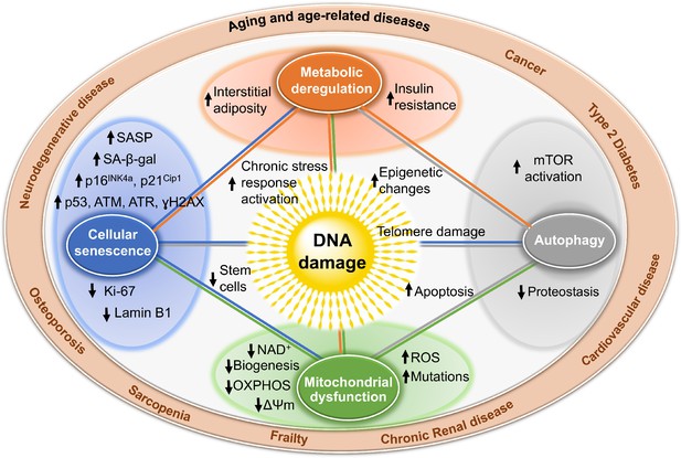
Mechanisms by which DNA impairment could promote aging.
Dna harm, including harm at telomeres (centre), one time detected, if not repaired, can interfere with replication or transcription, resulting in the activation of signaling events that change cell physiology. One effect of these signaling events is apoptosis, which while depleting important cells similar stalk cells or neurons may not be the near potent driver of aging. Dna damage can also result in mitochondrial dysfunction, impaired autophagy, metabolic changes, and the triggering of cellular senescence (modest circles). These alive just physiologically contradistinct cells are predicted to exist a more potent commuter of crumbling and disease. Endpoints used to measure these consequences of DNA damage are indicated with arrows in the larger circles. These outcomes are all interconnected in that mitochondrial dysfunction tin cause metabolic changes, impaired autophagy and proteostasis, more than DNA damage, and senescence. This creates a wheel of increasing harm and dysfunction, which can spread to other cells via SASP, that is likely the proximal crusade of aging and the diseases associated with it (outer circumvolve).
Boosted show to support the notion that genotoxic stress profoundly perturbs cellular homeostasis includes the following. Cells from patients with XP, CS, and AT have altered energy homeostasis, dumb mitophagy, and an increased mitochondrial membrane potential, implying accumulation of dysfunctional mitochondria and increased ATP and oxygen consumption (Valentin-Vega et al., 2022; Scheibye-Knudsen et al., 2022; Fang et al., 2022). Poly(ADP-ribose) polymerase ane (PARP1) is critical for the detection and repair of strand breaks. Persistent activation of PARP1 depletes cellular reserves of nicotinamide adenine dinucleotide (NAD+) (Finkel et al., 2009), a critical co-factor for many enzymes including sirtuins, which are a family of protein deacetylases and ADP-ribosyltransferases that broadly regulate factor expression and poly peptide stability. SIRT1 besides regulates mitochondrial biogenesis by deacetylating PGC-1α (peroxisome proliferator-activated receptor γ coactivator 1 α) (Finkel et al., 2009). SIRT1 activity is dramatically reduced in animal models of XP and CS due to persistent activation of PARP1 (Scheibye-Knudsen et al., 2022; Fang et al., 2022). Inhibition of PARP1 or supplementation with NAD+ precursors restore SIRT1 activity and improve mitochondrial homeostasis and cellular metabolism (Scheibye-Knudsen et al., 2022; Fang et al., 2022).
Is more Dna repair beneficial?
The gold standard for establishing a causal relationship betwixt 2 events (Deoxyribonucleic acid damage and aging) is to demonstrate that dumb repair accelerates aging, while improved repair slows aging. This is tricky with DNA repair as there are no drugs that stimulate Deoxyribonucleic acid repair, nor is it easy to meliorate DNA repair genetically. DNA repair mechanisms require the coordinated action of numerous proteins. Overexpression of just ane protein does non ever improve repair and, in fact, can be detrimental (Shaposhnikov et al., 2022). Nevertheless, in that location are some hints that longevity correlates with improved responses to genotoxic stress. In the nematode Caenorhabditis elegans, 40 single factor mutations have been described that increment lifespan past at least xx% and in all cases these mutations confer resistance to UV irradiation (Johnson et al., 2002). Overexpression of human MTH1, which prevents 8-oxoG accumulation, in mice protects against neurodegeneration (De Luca et al., 2008) and extends lifespan (De Luca et al., 2022). Enhancing ATM activity in a murine model of HGPS reduces progeroid features and extends lifespan (Qian et al., 2022). Interspecies comparisons take non definitively identified a correlation between Deoxyribonucleic acid repair chapters and lifespan (Austad, 2010; Cortopassi and Wang, 1996; Hart and Setlow, 1974), with a couple of exceptions. BER (but non NER) capacity is greater in cells from longer-lived rodents and non-human primates, than in shorter-lived species (Austad, 2010). Naked mole rats (lifespan 30+ years) have improved NER and BER efficiency relative to mice (lifespan 3 years) (Evdokimov et al., 2022). Many longer-lived species have increased expression or sequence optimization of fundamental regulators of genome stability (Keane et al., 2022; Tian et al., 2022) leading to improved DSB repair in longer-lived mammals (Tian et al., 2022). Cells of centenarians have improved Dna repair action and antioxidant capacity compared to non-centenarians (Chevanne et al., 2003; Franzke et al., 2022a). Calorie restriction (CR) is the most successful intervention to extend lifespan and/or health span in organisms ranging from yeast to mammals, including not-human being primates. CR has been demonstrated to decrease the abundance of DNA damaging reactive oxygen species (reviewed in Pamplona and Barja, 2006), thereby reducing oxidative Dna damage (reviewed in Heydari et al., 2007). Consistent with this is the observation that CR reduces transcriptional stress in Deoxyribonucleic acid repair defective mice (Vermeij et al., 2022). However, at that place is also prove that CR might amend DNA repair, including BER, NER, and NHEJ (studies in rodents thoroughly reviewed in Heydari et al., 2007). In humans, there is bear witness that CR (Matt et al., 2022), dietary micronutrients (Ames, 2010), chronic practise, and improved socialization of the elderly (Franzke et al., 2022b) can raise genome stability, which is ascribed to improved Dna repair capacity. However, more studies using robust measures of DNA repair capacity are needed. Collectively, there is abundant testify that more Dna repair at least correlates with improved healthspan and lifespan.
Conclusions
In that location is now sufficient and various testify to back up a denoting argument that DNA damage plays a causal function in aging. This includes environmental/iatrogenic sources of genotoxic stress as well as spontaneous/endogenous genotoxic stress. Deoxyribonucleic acid damage contributes to aging via cell democratic events such as causing apoptosis, which depletes functional cells such every bit neurons, and via cell non-autonomous mechanisms such as triggering senescence, which can negatively affect the function of neighboring, undamaged cells through their SASP. Downstream consequences of Dna damage impinge upon all of the other pillars of aging resulting in a state of self-perpetuating damage, which probable is the ultimate cause of aging. Despite these broad consequences of genotoxic stress, there is also evidence that these consequences tin can be modulated through approaches aimed at slowing crumbling, including caloric restriction, NAD+ supplementation, or ablating senescent cells. The field is still lacking tools to measure out Deoxyribonucleic acid lesions and DNA repair capacity that are accessible to the broader research community. Building such a tool kit would enable more precise conclusion of when (nether what circumstances) and where (in what organs) DNA damage truly drives aging. It also might open new opportunities in precision medicine, enabling fine tuning of DNA impairment and repair to, for case, better tumor ablation, slow the loss of irreplaceable cells, or optimize metabolism to promote repair.
References
-
Conference
Cancer treatment as an accelerated aging process: assessment, biomarkers, and interventions
American Society of Clinical Oncology Educational Book. American Social club of Clinical Oncology. Annual Meeting. pp. e516–e522.
- Google Scholar
Article and author information
Writer details
Funding
National Institutes of Health (P01 AG043376)
- Paul Robbins
- Laura Niedernhofer
National Institutes of Wellness (U01 ES029603)
- Laura Niedernhofer
National Institutes of Health (R56 AG059676)
- Laura Niedernhofer
National Institutes of Wellness (R01 AG063543)
- Paul Robbins
- Laura Niedernhofer
American Federation for Crumbling Inquiry (Irene Diamond Fund/American Federation for Aging Research Postdoctoral Transition Laurels)
- Matt Yousefzadeh
National Institutes of Health (U19 AG056278)
- Paul Robbins
- Laura Niedernhofer
The funders had no role in study design, data collection and interpretation, or the conclusion to submit the piece of work for publication.
Acknowledgements
We give thanks Mariah Witt for helpful comments. This work was supported past NIH grants P01 AG043376, U01 ES029603, R56 AG059676, R01 AG063543, and U19 AG056278. MJY was supported by the Irene Diamond Fund/American Federation for Aging Research Postdoctoral Transition Laurels.
Senior Editor
- Jessica Yard Tyler, Weill Cornell Medicine, United States
Reviewing Editor
- Matthew Simon, University of Rochester, U.s.a.
Publication history
- Received: September 9, 2022
- Accepted: January 15, 2022
- Version of Tape published: January 29, 2022 (version ane)
Copyright
© 2022, Yousefzadeh et al.
This article is distributed under the terms of the Artistic Commons Attribution License, which permits unrestricted apply and redistribution provided that the original author and source are credited.
Metrics
-
- thirteen,549
- Page views
-
- 963
- Downloads
-
- 27
- Citations
Article citation count generated by polling the highest count across the following sources: Crossref, PubMed Central, Scopus.
Download links
A two-function list of links to download the article, or parts of the commodity, in various formats.
Downloads (link to download the commodity as PDF)
Download citations (links to download the citations from this article in formats uniform with various reference managing director tools)
Open citations (links to open the citations from this article in various online reference manager services)

Can Damaged Dna Be Repaired,
Source: https://elifesciences.org/articles/62852
Posted by: gonzaleznathat.blogspot.com
0 Response to "Can Damaged Dna Be Repaired"
Post a Comment